Calcific Tendinopathy
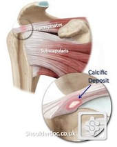
Calcific tendinopathy is a common condition, that occurs when calcium deposits in the shoulder tendons become painful.

It is most common between the ages of 30 - 50 years. Nobody knows why calcium is deposited in shoulder tendons, but in many cases it may be there for years and not cause any problems. Imaging (such as an x-ray or ultrasound scan) is required to confirm the diagnosis. In many cases, these calcific deposits can be managed non-‐surgically, however in some cases surgery may be required if conservative treatment measures do not provide adequate relief of symptoms.
Resorptive Calcific Tendinopathy
For unknown reasons, the body recognises the calcium and the area becomes inflamed as the body attempts to absorb, and break down the calcium. This is called "resorptive" calcific tendinopathy, and can be a painful condition. The pain can start spontaneously, or it may come on after a minor injury. The acute pain may last up to 2 weeks after which it may resolve on its' own, or may leave residual symptoms that require treatment.
For unknown reasons, the body recognises the calcium and the area becomes inflamed as the body attempts to absorb, and break down the calcium. This is called "resorptive" calcific tendinopathy, and can be a painful condition. The pain can start spontaneously, or it may come on after a minor injury. The acute pain may last up to 2 weeks after which it may resolve on its' own, or may leave residual symptoms that require treatment.

Treatment Options
1. Do nothing.
Once the initial pain has settled, shoulder function can recover spontaneously over several weeks without the need for any treatment, beyond basic pain relief medications.
2. Ultrasound-Guided Injection.
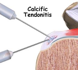
An injection of anaesthetic and corticosteroid (anti-‐inflammatory) performed under the guidance of ultrasound imaging can provide effective relief for those cases where pain is severe and disabling. If the calcium deposits are large, causing interference with normal shoulder movement and ‘impingement’ type symptoms, they can also be treated with a procedure called fenestration (needling of the calcific deposits performed using local anaesthetic), or barbotage (injection of saline (salt water), local anaesthetic and corticosteroid (anti-‐inflammatory) into the calcific deposits). Both of these procedures are done under ultrasound guidance. This can be repeated 2-3 times over a period of several months if symptoms are persistent.
3. Surgery
If the calcium deposits are large and troublesome, surgical excision may be required. Usually, surgery is only recommended if the other procedures (analgesics, anti-‐inflammatory medications and injection procedures) are unsuitable or have been unsuccessful.